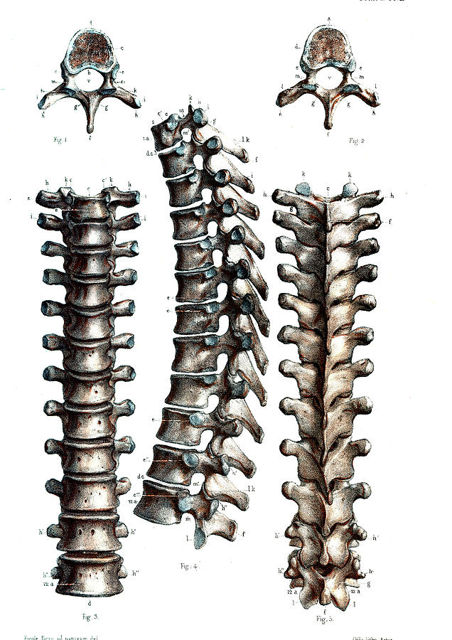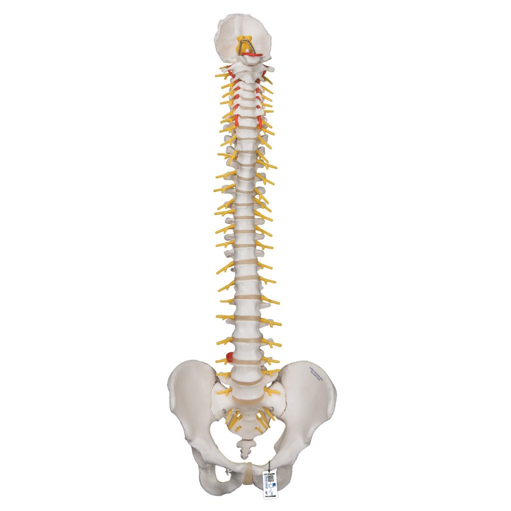


These structures form the vertebral canal, which contains the spinal cord, spinal nerves, and epidural space. Two pedicles arise on the posterolateral aspects of each vertebra and fuse with the two laminae to encircle the vertebral foramen1 ( Figure 2A, Figure 2B). This holds true for all vertebrae except C1. VertebraeĪ typical vertebra consists of a vertebral arch posteriorly and a body anteriorly. The vertebral column and the curvatures of the adult spine, lateral view. The secondary curvatures are convex anteriorly and augment the flexibility of the spine.įIGURE 1. The cervical and lumbar lordoses are secondary curvatures that develop after birth as a result of extension of the head and lower limbs when standing erect. These curvatures persist through adulthood as the thoracic and sacral curves. In the embryonic period, the spine curves into a C shape, forming two primary curvatures with their convex aspect directed posteriorly. The vertebral column consists of 33 vertebrae (7 cervical, 12 thoracic, 5 lumbar, 5 sacral, and 4 coccygeal segments) ( Figure 1). It is important, therefore, that the anesthesiologist be able to develop a three-dimensional mental image of the structures comprising the vertebral column. Pertinent to regional anesthesia, the vertebral column serves as the landmark for a wide variety of regional anesthesia techniques. Its four primary functions are protection of the spinal cord, support of the head, provision of an attachment point for the upper extremities, and transmission of weight from the trunk to the lower extremities. The vertebral column forms part of the axis of the human body, extending in the midline from the base of the skull to the pelvis. Orebaugh and Hillenn Cruz Eng INTRODUCTION He has received numerous international awards as an orthopedic trainee including an RCSI/Ethicon bursary, BOA/Zimmer Biomet travelling fellowship, SOFCOT/ARMO foreign graduate award and the Mark Paterson/EFORT travelling fellowship.Steven L.

Having completed his training as an orthopedic surgeon in the Republic of Ireland, he is the author of several publications on orthopedic and spinal research topics. Derek Thomas Cawley is a Spinal Fellow at Bordeaux University Hospital.

He also serves on the editorial boards of the European Spine Journal, The Spine, and The French Journal of Orthopedic and Traumatology Surgery.ĭr. He has been a member of several outstanding societies such as the French Medical College of Anatomy since 1989, and the European Cervical Spine Research Society since 2003. Vital has a special interest in spinal deformities (with particular emphasis on sagittal balance) and in cervical spine surgery (cervical prostheses and myelopathy). He also earned the national specialized Diploma in Sports Medicine (CES).Īs a spine surgeon, Dr. In the same year, he became Senior Registrar of the Department run by Prof. In 1981 he was appointed Instructor of Anatomy and Organogenesis as well as Intern in Orthopedic Surgery and Traumatology. Moreover, he earned an MD in human biology in the field of anatomy. Upon completing his residency in Bordeaux in 1980, he received the Gold Medal Award of Surgery. Since 1989, Jean Marc Vital has been an Intern and University Professor of orthopedic and traumatology surgery at the University of Medicine of Bordeaux, as well as Head of the Department of Spinal Diseases and Director of the Anatomy Laboratory at the Paul Broca faculty. New histological and chemical findings on the intervertebral disc, as well as detailed descriptions of the aponeuroses and fasciae, are also provided.īringing together the experience of several experts from the well-known French school, this book offers a valuable companion for skilled experts and postgraduate students in various fields: orthopedic surgery, neurosurgery, physiotherapy, rheumatology, musculoskeletal therapy, rehabilitation, and kinesiology. the 3D study of vertebral movements using the “EOS system,” which makes it possible to define an equilibrium of posture and its limits. The respective chapters review in depth all sections of the vertebral column and offer new insights, e.g. The dynamic anatomy of the living subject is viewed using the latest technologies, opening new perspectives to elucidate the pathology of the spine and improve spinal surgery. This richly illustrated and comprehensive book covers a broad range of normal and pathologic conditions of the vertebral column, from its embryology to its development, its pathology, its dynamism and its degeneration.


 0 kommentar(er)
